Dodo and other extinct birds Plate 33

Description:
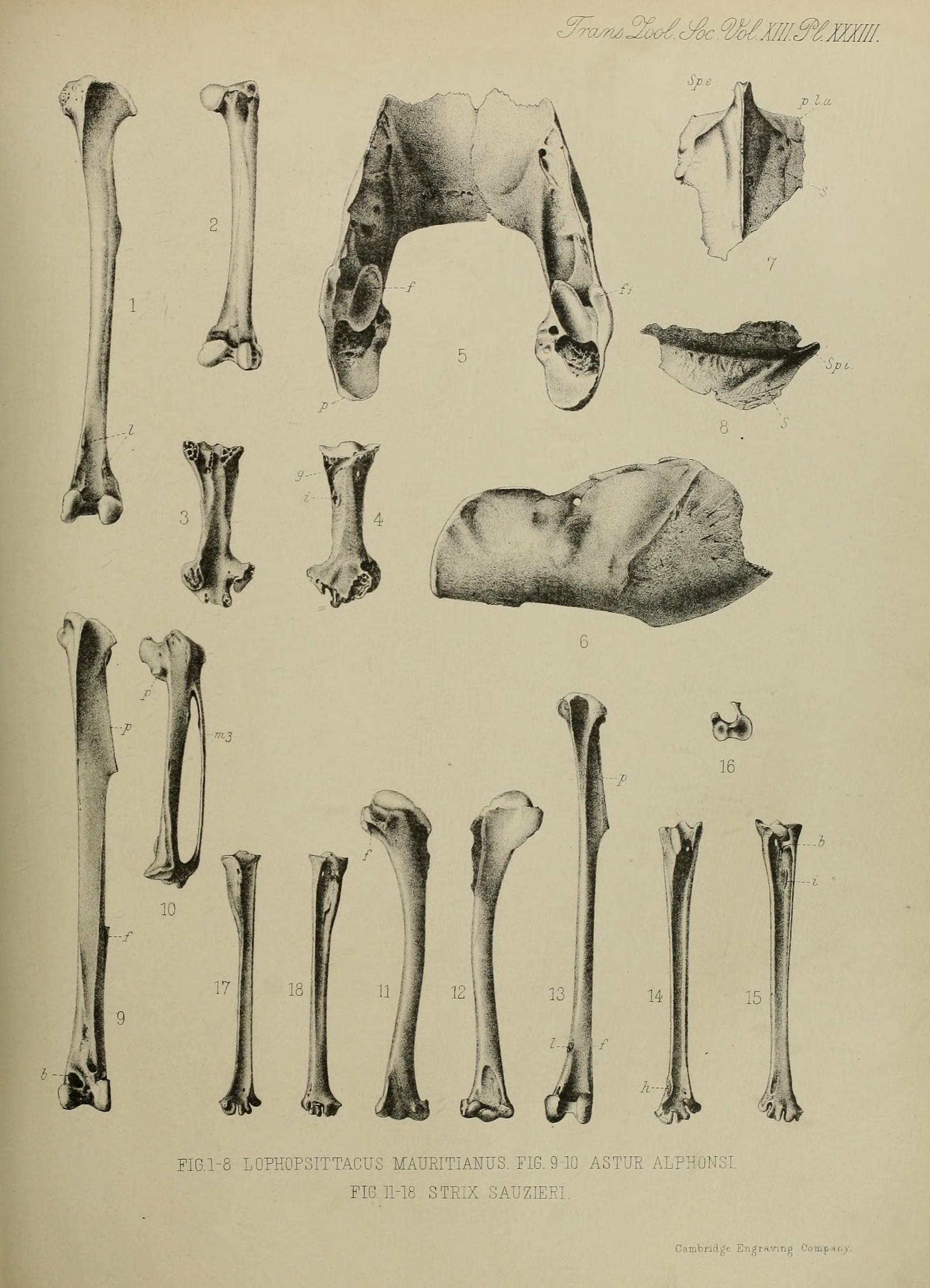
Plate XXXIII from Edward Newton and Hans Gadow's On additional bones of the Dodo and other extinct birds of Mauritius obtained by Mr. Théodore Sauzier. 1–8: Lophopsittacus mauritianus 1 Left tibia, front view. l: attachments of the transverse ligament across the long extensor tendons. 2 Right femur, posterior view. 3 Left metatarsus, plantar surface. 4 Left metatarsus, dorsal surface, g: groove for the tendon of the musculus extensor digitorum; i: insertion of the tendon of the m. tibialis anticus. 5 Dorsal view of the underjaw. p: posterior angle; f: facet for the quadrate; fi: additional facet for the jugal process of the quadrate. 6 Lateral view of the right jaw. 7 Ventral view of sternum. Sp.e.: spina externa sterni; p.l.a.: anterior lateral process; S: lateral line of m. subclavius. 8 Right lateral view of sternum. Sp.i.: spina externa sterni ; S: median line of m. subclavius. 9–10: Circus maillardi 9 Left tibia, front view. f: rest of fibula; b: bony bridge over the tendon of the m. extensor digitorum; p: peroneal crest. 10 Left metacarpals, lateral view. p: articular facet of the pollex; m3: metacarpale III 11–18: Mascarenotus sauzieri 11 Inner view of humerus. f: pneumatic foramen. 12 Outer view of humerus. 13 Left tibia, front view. p: peroneal crest; f: distal portion of fibula ; l: attachment of transverse ligament. 14 Posterior view of right tarso-metatarsus. h: bony bridge across the tendon of the m. tibialis anticus. 15 Anterior view of right tarso-metatarsus. b: facet for the hallux's metatarsal; i: insertion of the m. tibialis anticus. 16 Proximal end of the tarsus. 17 Posterior view of the small pair of tarso-metatarsals. 18 Anterior view of the small pair of tarso-metatarsals.
Included On The Following Pages:
- Life
- Cellular
- Eukaryota (eukaryotes)
- Opisthokonta (opisthokonts)
- Metazoa (animals)
- Bilateria
- Deuterostomia (deuterostomes)
- Chordata (Chordates)
- Vertebrata (vertebrates)
- Gnathostomata (jawed fish)
- Osteichthyes (bony fish)
- Sarcopterygii (Lobe-finned fishes)
- Tetrapoda (terrestrial vertebrates)
- Amniota (amniote)
- Reptilia (Reptiles)
- Diapsida (diapsid)
- Archosauromorpha (archosauromorph)
- Archosauria (archosaur)
- Dinosauria (dinosaurs and birds)
- Saurischia (saurischian)
- Theropoda (theropods)
- Tetanurae (tetanuran theropod)
- Coelurosauria (coelurosaur)
- Maniraptoriformes
- Maniraptora (maniraptoran)
- Aves (birds)
- Ornithurae
- Neornithes
- Neognathae
- Neoaves
- landbirds
- Accipitriformes (raptors)
- Accipitridae (hawks, eagles, and relatives)
- Circus
- Circus maillardi (Reunion Harrier)
- Paraves
This image is not featured in any collections.
Source Information
- license
- cc-licenses-publicdomain
- original
- original media file
- visit source
- partner site
- Wikimedia Commons
- ID