Illacme plenipes anatomy Fig. 2–5

Description:
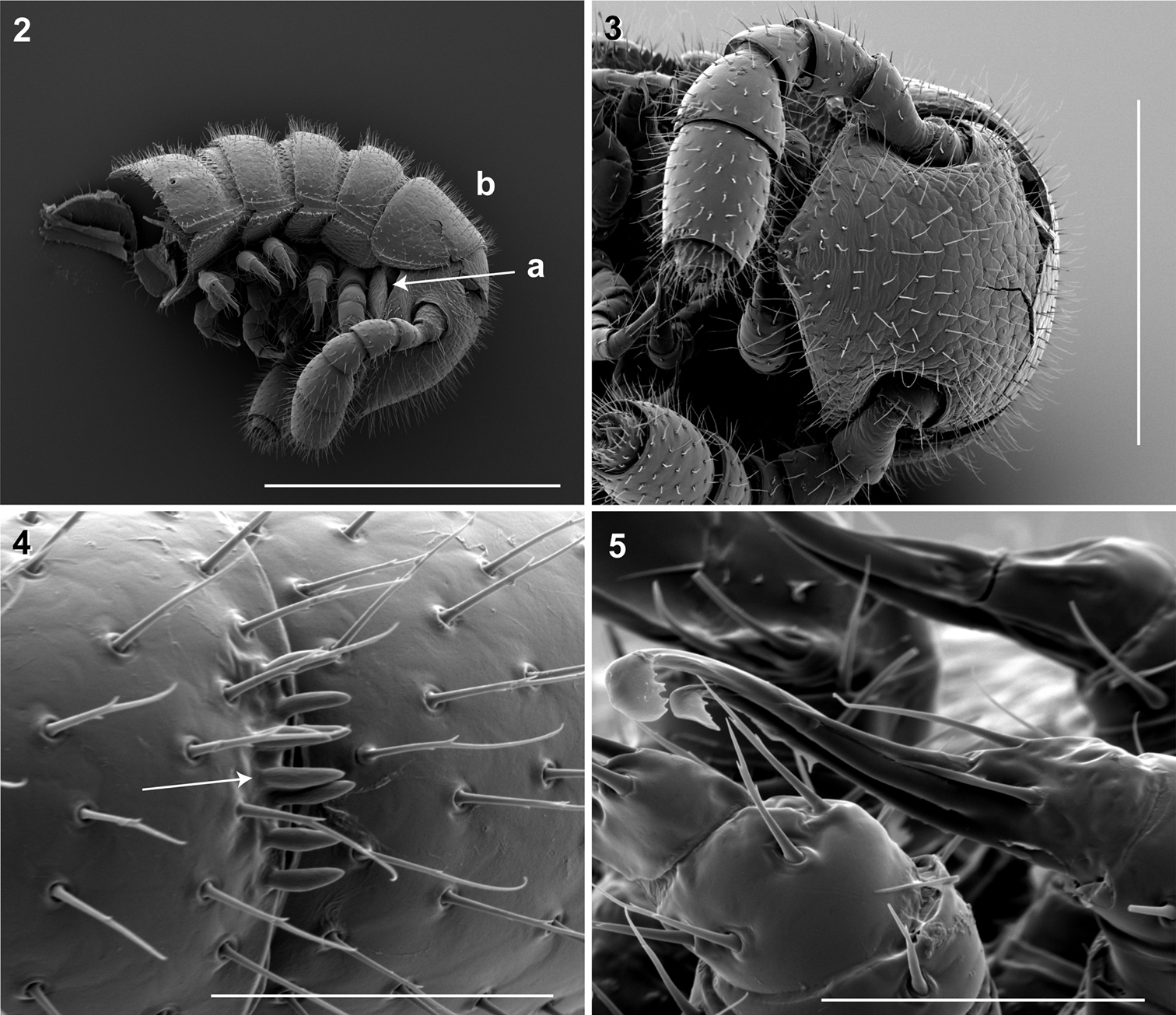
2: Lateral (right) view of head and segments 1–5 (♂). a Lateral opening apparent between gnathochilarium and head capsule; gnathochilarium, mandible and head capsule noticeably separate at base, 1/3 head length distally from mandibular joint b Collum not covering head, with straight cephalic edge, gradually tapering laterally. Scale bar 0.5 mm. 3: Ventral view of head, antennae and segments 1 – 5 (♂).Scale bar 0.3 mm. 4: Lateral (right) view of antennomeres 5, 6 (♂).Arrow, small basiconic sensilla (Bs2) in cluster of 7 or 8 oriented apical dorsally (retrolaterally) in slight depression on antennomeres 5, 6. Scale bar 0.05 mm. 5 Oblique (right) view of right posterior gonopod (♂).Posterior gonopodal podomere 6 divided, comprising a bundle of 3 stylus-shaped articles. Scale bar 0.05 mm.
Included On The Following Pages:
- Life
- Cellular
- Eukaryota (eukaryotes)
- Opisthokonta (opisthokonts)
- Metazoa (animals)
- Bilateria
- Protostomia (protostomes)
- Ecdysozoa (ecdysozoans)
- Arthropoda (arthropods)
- Myriapoda (myriapods)
- Diplopoda (millipedes)
- Siphonophorida
- Galbella ennediana
- Galbella regina
- Galbella occidentalis
- Panarthropoda
This image is not featured in any collections.
Source Information
- license
- cc-by-3.0
- copyright
- Paul E. Marek, William A. Shear, Jason E. Bond
- creator
- Paul E. Marek, William A. Shear, Jason E. Bond
- original
- original media file
- visit source
- partner site
- Wikimedia Commons
- ID