Giardia 1

Description:
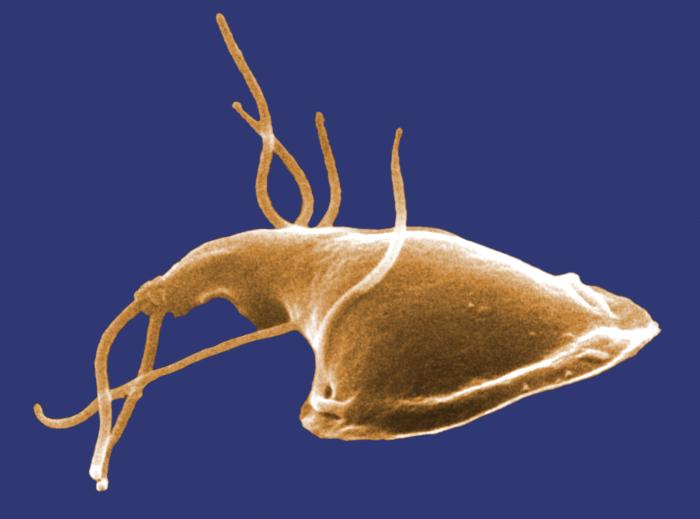
Summary[edit] Description: English: ID#: 11649 Description: This digitally-colorized scanning electron micrograph (SEM) depicted the dorsal (upper) surface of a Giardia protozoan that had been isolated from a rat’s intestine. Some of the identifying morphologic characteristics include pairs of thread-like flagella that facilitate motility, and a ventolateral flange that appears as a “ruffle” around the anterior portion of the organism. Pairs of flagella seen here include an anterior, posterior-lateral, and caudal pairs. The protozoan Giardia causes the diarrheal disease called giardiasis. Giardia species exist as free-swimming (by means of flagella) trophozoites, and as egg-shaped cysts. It is the cystic stage, which facilitates the survival of these organisms under harsh environmental conditions. The cyst is considered the infective form, and disease is often transmitted by drinking contaminated water. As depicted in these SEMs, in the intestine, cysts are stimulated to liberate trophozoites. Cysts can be shed in fecal material, and can, thereafter, remain viable for several months in appropriate environmental conditions. Cysts can also be transferred directly from person-to-person, as a result of poor hygiene. Dr. Stan Erlandsen; Dr. Dennis Feely. Date: 1982. Source: http://phil.cdc.gov/PHIL_Images/11649/11649_lores.jpg. Author: Dr. Stan Erlandsen; Dr. Dennis Feely,Center for Disease Control.
Included On The Following Pages:
- Life
- Cellular
- Eukaryota (eukaryotes)
- Excavates (excavates)
- Metamonada (metamonad)
- Fornicata
- Diplomonadida
- Hexamitidae
- Cryptoplus ferrugineus
- Giardia
This image is not featured in any collections.
Source Information
- license
- cc-licenses-publicdomain
- creator
- Dr. Stan Erlandsen; Dr. Dennis Feely,Center for Disease Control
- source
- http://phil.cdc.gov/PHIL_Images/11649/11649_lores.jpg
- original
- original media file
- visit source
- partner site
- Wikimedia Commons
- ID