Image of Hitobia

Description:
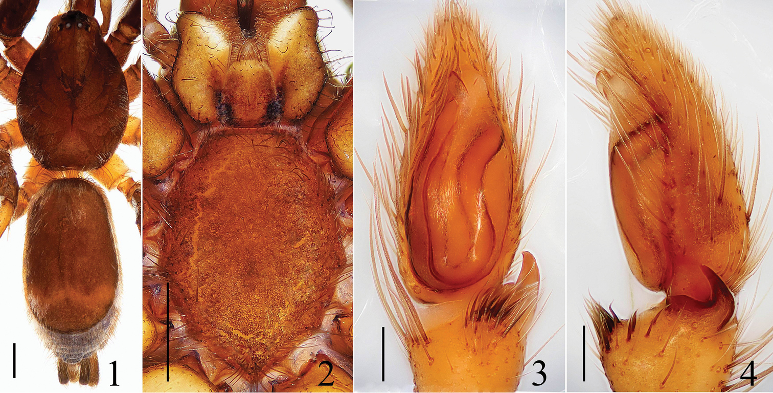
Figures 1–4.Hitobia tengchong sp. n. 1 male body, dorsal view 2 prosoma, ventral view 3 male palp, ventral view 4 male palp, retrolateral view. Scale bars: 0.5 mm (1–2); 0.1 mm (3–4).
Included On The Following Pages:
- Life
- Cellular
- Eukaryota (eukaryotes)
- Opisthokonta (opisthokonts)
- Metazoa (animals)
- Bilateria
- Protostomia (protostomes)
- Ecdysozoa (ecdysozoans)
- Arthropoda (arthropods)
- Chelicerata (chelicerates)
- Arachnida (arachnids)
- Araneae (spiders)
- Opisthothelae
- Araneomorphae
- Entelegynae
- Retrolateral tibial apophysis
- Gnaphosidae (ground spiders)
- Hitobia
- Hitobia tengchong
- Panarthropoda
This image is not featured in any collections.
Source Information
- license
- cc-by-3.0
- copyright
- Cheng Wang, Xian-Jin Peng
- bibliographic citation
- Wang C, Peng X (2014) Three species of Hitobia Kamura, 1992 (Araneae, Gnaphosidae) from south-west China ZooKeys (464): 25–34
- original
- original media file
- visit source
- partner site
- Zookeys
- ID