Image of Xestoblatta

Description:
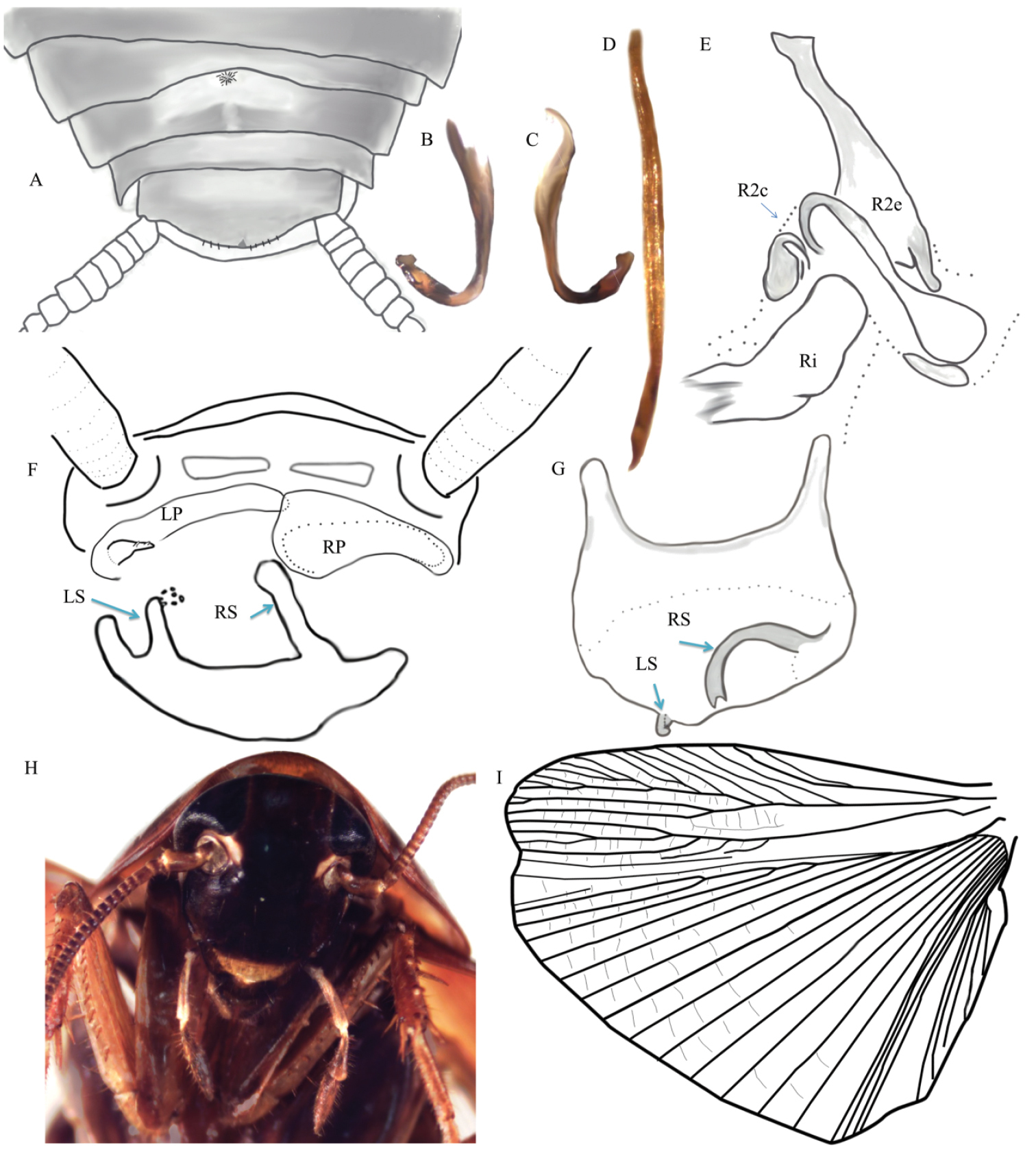
Figure 9.Xestoblatta berenbaumae sp. n. A Dorsal view of abdomen showing the simple tergal gland (DECBA2023) B, C Hooked left phallomere D Ventral medial phallomere (L2vm) E Right phallomere. R2e – external sclerite, R2i – internal sclerite, R2c – cleft sclerite F Posterior view of abdomen showing paraprocts and subgenital plate. RS-right stylus, LS-left stylus with small translucent ball at tip, LP-left paraproct reduced and specialized with polydentate spine, RP-unspecialized right paraproct. Illustration is a composite of multiple individuals G Dorsal view of sub-genital plate (DECBA1967) H Head of adult male I Hindwing (DECBA0801). Photos and illustrations contributed by Kayla Kaplan and Dominic A. Evangelista.
Included On The Following Pages:
- Life
- Cellular
- Eukaryota (eukaryotes)
- Opisthokonta (opisthokonts)
- Metazoa (animals)
- Bilateria
- Protostomia (protostomes)
- Ecdysozoa (ecdysozoans)
- Arthropoda (arthropods)
- Pancrustacea
- Hexapoda (hexapods)
- Insecta (insects)
- Pterygota (winged insects)
- Neoptera
- Polyneoptera
- Dictyoptera
- Blattodea (cockroaches and termites)
- Blaberoidea
- Ectobiidae (german cockroach family)
- Xestoblatta
- Xestoblatta berenbaumae
- Panarthropoda
This image is not featured in any collections.
Source Information
- license
- cc-by-3.0
- copyright
- Dominic A. Evangelista, Kimberly Chan, Kayla L. Kaplan, Megan M. Wilson, Jessica L. Ware
- bibliographic citation
- Evangelista D, Chan K, Kaplan K, Wilson M, Ware J (2015) The Blattodea s.s. (Insecta, Dictyoptera) of the Guiana Shield ZooKeys (475): 37–87
- original
- original media file
- visit source
- partner site
- Zookeys
- ID