Image of Argyrodiaptomus Brehm 1933

Description:
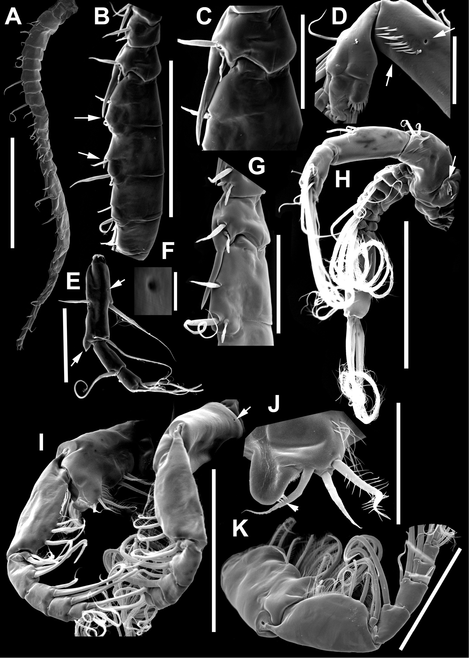
Figure 5.Argyrodiaptomus azevedoi male, SEM photographs. A Geniculate right antennule (500 µm) B Segments 12–17 of A1R (300 µm) C Segments 13–14 of A1R (100 µm) D Enp of A2, showing pore and spinular ornamentation (50 µm) E Segments 20–22 of A1R (100 µm) F Inset showing pore on segment 20 of A1R (5 µm) G Segments 12–16 of A1R (100 µm) H A2 (200 µm) I Maxillipeds (200 µm) J Distal endite of maxilliped (50 µm) K Maxilliped (200 µm).
Included On The Following Pages:
- Life
- Cellular
- Eukaryota (eukaryotes)
- Opisthokonta (opisthokonts)
- Metazoa (animals)
- Bilateria
- Protostomia (protostomes)
- Ecdysozoa (ecdysozoans)
- Arthropoda (arthropods)
- Pancrustacea
- Multicrustacea (typical crustaceans)
- Copepoda (copepods)
- Calanoida (Calanoid copepods)
- Diaptomidae
- Argyrodiaptomus
- Argyrodiaptomus azevedoi
- Hexanauplia
- Panarthropoda
This image is not featured in any collections.
Source Information
- license
- cc-by-3.0
- copyright
- Gilmar Perbiche-Neves, Geoffrey Allan Boxshall, Daniel Previattelli, Marcos Gomes Nogueira, Carlos Eduardo Falavigna da Rocha
- bibliographic citation
- Perbiche-Neves G, Boxshall G, Previattelli D, Nogueira M, da Rocha C (2015) Identification guide to some Diaptomid species (Crustacea, Copepoda, Calanoida, Diaptomidae) of “de la Plata” River Basin (South America) ZooKeys (497): 1–111
- original
- original media file
- visit source
- partner site
- Zookeys
- ID