Cicerina debrae

Description:
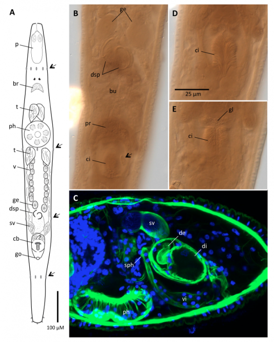
Cicerina debrae n. sp. Anterior end is to the left in Fig C and to the top in the remaining figures. A. General anatomy; arrows, adhesive belts; br, brain; cb, copulatory bulb; dsp, ductus spermaticus; ge, germarium; go, common gonopore; ph, pharynx; sv, seminal vesicle; t, testes; v, vitellaria. B. Bright-field micrograph of a specimen from a resinslide preparation, showing genital region with paired germaria, ducti spermatici with flanges where they enter the bursal tissue (bu), proximal prostatic region of copulatory bulb (pr), and distal portion of copulatory bulb with cirrus (ci) and matrix-cell nucleus (arrow). C. CLSM stack (Alexa488/phalloidi?green; Hoechst 3334?blue) of genital region, lateral view showing seminal vesicle, proximal region of copulatory bulb with invaginated internal ductus ejaculatorius (de), distal (di) region of copulatory bulb with separate muscle layer, and vagina interna (vi) ending in a sphincter (sph) at the bursal tissue. D, E. Brightfield micrographs of same specimen as B, more-dorsal and more-ventral (respectively) focal planes of the cirrus, showing arrangement of spines, and hemi-circular wreath of gland cells (gl) located at the base of the cirrus.
Included On The Following Pages:
- Life
- Cellular
- Eukaryota (eukaryotes)
- Opisthokonta (opisthokonts)
- Metazoa (animals)
- Bilateria
- Protostomia (protostomes)
- Spiralia (spiralians)
- Platyhelminthes (flatworms)
- Rhabdocoela
- Cicerinidae
- Cicerina
- Cicerina debrae
- Rhabditophora
This image is not featured in any collections.
Source Information
- license
- cc-by-nc-sa-4.0
- copyright
- WoRMS Editorial Board
- contributor
- Tucker et al., 2014
- original
- original media file
- visit source
- partner site
- World Register of Marine Species
- ID