Summary[edit] Description: English: Gram stain of Methylobacterium fujisawaense appear as negative rods under bright field microscopy x1000. Date: 14 October 2013, 13:21:43. Source: Own work. Author: Dr. Sahay.
Summary[edit] Description: English: Phages of Ochrobactrum sp. POC9. Characteristics of the virions and plaques of the vB_OspP_OH phage (unclassified Podoviridae, jumbophage). The scale bars in the transmission electron microscopy (TEM) images represent 50 nm. The phage plaques are presented in the insets on panel (plaques of vB_OspP_OH). The scale bars in the insets represent 1 mm. Date: 18 March 2020. Source: Fig. 1b at https://www.mdpi.com/1422-0067/21/6/2096/htm Identification and Characterization of the First Virulent Phages, Including a Novel Jumbo Virus, Infecting Ochrobactrum spp.; MDPI Int. J. Mol. Sci. 2020, 21(6), doi:10.3390/ijms21062096. Author: Przemyslaw Decewicz, Piotr Golec, Mateusz Szymczak, Monika Radlinska, Lukasz Dziewit. Other versions:.
Summary[edit] Description: English: The bacterium Bradyrhizobium japonicum strain USDA 110 streaked to single colonies on Tryptone-Yeast Extract (TY) agar. Date: 31 July 2010. Source: Own work. Author: Ninjatacoshell.
Summary[edit] Description: English: The root system of a 4-week-old Medicago italica. Date: 20 July 2009. Source: Own work. Author: Ninjatacoshell. : This is a retouched picture, which means that it has been digitally altered from its original version. Modifications: I used GIMP 2.6.3 to blacken the background and to increase the saturation by 50%..
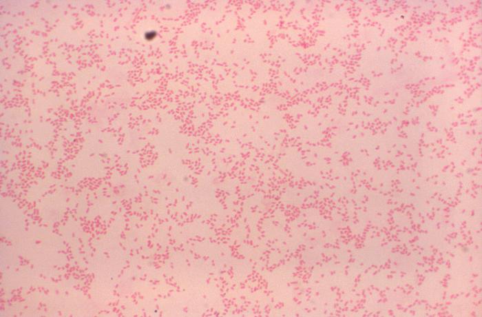
Summary[edit] Description: English: Processed using the Gram-stain method, this photomicrograph revealed the presence of numerous Gram-negative, coccobacillus, Brucella suis bacteria. Date: 1977. Source: https://phil.cdc.gov/Details.aspx?pid=19255. Author: CDC/ Dr. W.A. Clark.
Summary[edit] Description: English: Oxytropis dinarica ssp. dinarica var. macrocarpa leg P.Cikovac Nodule on Taproot. Date: 25 July 2015. Source: Own work. Author: P.Cikovac.
Summary[edit] Description: English: Methylobacterium fujisawaense pink-pigmented bacterial colonies growing on R2A after 5 days of incubation at 30° Celsius (front view). Date: 14 October 2013, 13:06:25. Source: Own work. Author: Dr. Sahay.
Summary[edit] Description: English: Closeup of a root nodule on a Medicago plant, inoculated with Sinorhizobium meliloti. The nodule has been cut in half and demonstrates the four zones of an indeterminate nodule. Date: 22 July 2009. Source: Own work. Author: Ninjatacoshell.
Description: ID#:1902 Brucella melitensis colonies. Brucella spp. Colony Characteristics: A. Fastidious, usually not visible at 24h. B. Grows slowly on most standard laboratory media (e.g. sheep blood, chocolate and trypticase soy agars). Pinpoint, smooth, entire translucent, non-hemolytic at 48h. Content Providers(s):CDC/ Courtesy of Larry Stauffer, Oregon State Public Health Laboratory Creation Date:2002 Copyright Restrictions:None - This image is in the public domain and thus free of any copyright restrictions. As a matter of courtesy we request that the content provider be credited and notified in any public or private usage of this image. Source: : This media comes from the Centers for Disease Control and Prevention's Public Health Image Library (PHIL), with identification number #1902. Note: Not all PHIL images are public domain; be sure to check copyright status and credit authors and content providers. العربية | Deutsch | English | македонски | slovenščina | +/−. Author: This file is lacking author information. Permission(Reusing this file): PD.
Summary[edit] Description: English: The bacterium Rhizobium tropici strain BR816 streaked to single colonies on Tryptone-Yeast Extract (TY) agar. Português: Bactéria Rhizobium tropici estriada em placa de ágar Extrato de Levedura Triptona (LT). Date: 31 July 2010. Source: Own work. Author: Ninjatacoshell. Other versions: Derivative works of this file: Rhizobium tropici strain BR816 on TY agar-2.JPG.
Summary[edit] Description: English: The bacterium Sinorhizobium fredii strain USDA257 streaked to single colonies on Tryptone-Yeast Extract (TY) agar. Date:. Source: Own work. Author: Ninjatacoshell.
Summary[edit] Description: English: The root nodules of a 4-week-old Medicago italica inoculated with Sinorhizobium meliloti. Date: 20 July 2009. Source: Own work. Author: Ninjatacoshell. : This is a retouched picture, which means that it has been digitally altered from its original version. Modifications: I used GIMP 2.6.3 to blacken the background and to increase the saturation by 50%..
Summary[edit] Description: English: The bacterium Sinorhizobium meliloti strain Rm1021 streaked to single colonies on Tryptone-Yeast Extract (TY) agar. Date: 31 July 2010. Source: Own work. Author: Ninjatacoshell.
Summary[edit] Description: English: This Gram-stained photomicrograph depict numerous Gram-negative Brucella canis bacteria. B. canis is known to cause brucellosis in dogs and cats. Date: 1977. Source: http://phil.cdc.gov/PHIL_Images/15243/15243_lores.jpg. Author: Department of Health and Human Services.


