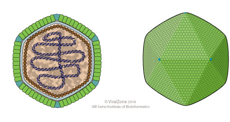
ViralZone, SIB Swiss Institute of Bioinformatics, see https://viralzone.expasy.org/ bottom right and Commons:Deletion requests/Files uploaded by Ernsts
Wikimedia Commons
Kenta Okamoto, Naoyuki Miyazaki, Hemanth K. N. Reddy, Max F. Hantke, Filipe R. N. C. Maia, Daniel S. D. Larsson, Chantal Abergel, Jean-Michel Claverie, Janos Hajdu, Kazuyoshi Murata, and Martin Svenda
Wikimedia Commons
Summary[edit] Description: English: Images of cryo-frozen Melbournevirus (family: Marseilleviridae) particles (left and center) and enlarged diagram of structure near a vertex. Black arrows indicate Large Dense Bodies. White arrows indicate lipid bilayer. Date: 17 May 2017. Source: from figure 2 of http://www.biorxiv.org/content/biorxiv/early/2017/05/17/139097.full.pdf Cryo-EM of a Marseilleviridae virus particle reveals a large internal microassembly. . Author: Kenta Okamoto, Naoyuki Miyazaki, Hemanth K. N. Reddy, Max F. Hantke, Filipe R. N. C. Maia, Daniel S. D. Larsson, Chantal Abergel, Jean-Michel Claverie, Janos Hajdu, Kazuyoshi Murata, and Martin Svenda.
Summary[edit] Description: English: Ultrathin-section TEM imaging of Noumeavirus-infected Acanthamoeba cells. (a) 10 min pi, virions have been engulfed and are in vacuoles. (b) 20 min pi, virions in vacuoles exhibit holes (black arrows) in their external translucent shell and appear more spherical. (c) 30 min pi, virions have entirely lost their external shell and appear as spherical electron-dense nucleoids, which can be seen in vacuoles as well as in the cell cytoplasm (and Fig. 9, black arrows). Scale bar, 100 nm. (d) 1 h pi, electron-dense tubular structures (black arrowheads) appear in the cytoplasm. (e) 4 h pi viral factories (VF) settle in the cell cytoplasm next to the nucleus and the cell organelles are pushed at their periphery (scale bar, 2 μm). The new virions are assembled and the electron-dense mature (white arrows) and immature (white arrowheads) virions are scattered in the same VF. Inset: immature and mature virions inside the VF along with tubular structures (black arrowheads). Scale bar, 200 nm. (f) 7 h pi, the newly synthesized virions are gathered into vacuoles inside the cytoplasm before being released in the extracellular environment (scale bar, 500 nm). Date: 21 April 2017. Source: https://media.springernature.com/full/springer-static/image/art%3A10.1038%2Fncomms15087/MediaObjects/41467_2017_Article_BFncomms15087_Fig1_HTML.jpg (contrast enhanced) at https://www.nature.com/articles/ncomms15087. Author: Elisabeth Fabre, Sandra Jeudy, Sébastien Santini, Matthieu Legendre, Mathieu Trauchessec, Yohann Couté, Jean-Michel Claverie, Chantal Abergel. Other versions: .
Summary[edit] Description: English: Ultrathin-section TEM imaging of Noumeavirus-infected Acanthamoeba cells. (a) 10 min pi, virions have been engulfed and are in vacuoles. (b) 20 min pi, virions in vacuoles exhibit holes (black arrows) in their external translucent shell and appear more spherical. (c) 30 min pi, virions have entirely lost their external shell and appear as spherical electron-dense nucleoids, which can be seen in vacuoles as well as in the cell cytoplasm (and Fig. 9, black arrows). Scale bar, 100 nm. (d) 1 h pi, electron-dense tubular structures (black arrowheads) appear in the cytoplasm. (e) 4 h pi viral factories (VF) settle in the cell cytoplasm next to the nucleus and the cell organelles are pushed at their periphery (scale bar, 2 μm). The new virions are assembled and the electron-dense mature (white arrows) and immature (white arrowheads) virions are scattered in the same VF. Inset: immature and mature virions inside the VF along with tubular structures (black arrowheads). Scale bar, 200 nm. (f) 7 h pi, the newly synthesized virions are gathered into vacuoles inside the cytoplasm before being released in the extracellular environment (scale bar, 500 nm). Date: 21 April 2017. Source: https://media.springernature.com/full/springer-static/image/art%3A10.1038%2Fncomms15087/MediaObjects/41467_2017_Article_BFncomms15087_Fig1_HTML.jpg at https://www.nature.com/articles/ncomms15087. Author: Elisabeth Fabre, Sandra Jeudy, Sébastien Santini, Matthieu Legendre, Mathieu Trauchessec, Yohann Couté, Jean-Michel Claverie, Chantal Abergel. Other versions: .
Fábio P. Dornas, Felipe L. Assis, Sarah Aherfi, Thalita Arantes, Jônatas S. Abrahão, Philippe Colson and Bernard La Scola
Wikimedia Commons
All images in this article were uploaded in the JPEG format even though it consists of non-photographic data. This information could be stored more efficiently or accurately in the PNG or SVG format. If possible, please upload a PNG or SVG version of this image without compression artifacts, derived from a non-JPEG source (or with existing artifacts removed). After doing so, please tag the JPEG version with {{Superseded|NewImage.ext}} and remove this tag. This tag should not be applied to photographs or scans. For more information, see {{BadJPEG}}. Summary[edit] Description: English: Genome architecture and synteny of some marseilleviridae. Date: 10 March 2016. Source: figure 1 of http://www.mdpi.com/1999-4915/8/3/76/htm. Author: Fábio P. Dornas, Felipe L. Assis, Sarah Aherfi, Thalita Arantes, Jônatas S. Abrahão, Philippe Colson and Bernard La Scola.


