Summary[edit] Description: English: A silver stain of Legionella pneumophila, the bacteria that causes Legionellosis. Although I got this image from a commercial website it is clearly labeled as from the CDC. This website routinely uses images from Wikipedia, which is a good thing, so no issue should be taken with using an presumably public domain image from their website. Date: 28 May 2010. Source: www.usmlerx.com. Author: William Cherry.
Summary[edit] Description: English: Colorized scanning electron micrograph (SEM) with moderately-high magnification of 5000X, depicting a large grouping of Gram-negative Legionella pneumophila bacteria. Русский: Цветная электросканограмма, увеличение х5000. В поле зрения - Legionella pneumophila. Date: 2009. Source: : This media comes from the Centers for Disease Control and Prevention's Public Health Image Library (PHIL), with identification number #11148. Note: Not all PHIL images are public domain; be sure to check copyright status and credit authors and content providers. English | Slovenščina | +/−. Author: Janice Haney Carr; provided by CDC/ Margaret Williams, PhD; Claressa Lucas, PhD;Tatiana Travis, BS.
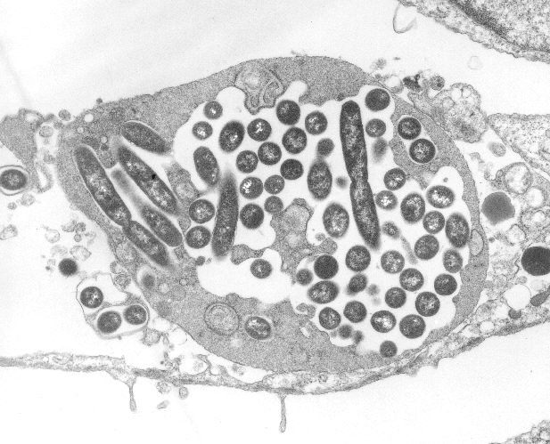
Summary[edit] Description: English: TEM image of cell infected with a number of Legionella pneumophila bacteria. Date: 19 October 2011. Source: TEM image of infected phagocytic cell Previously published: No. Author: Clares back.
Description: ID#: 934 Legionella pneumophila multiplying inside a cultured human lung fibroblast. Multiple intracellular bacilli, including dividing bacilli, are visible in longitudinal and cross section. Transmission electron micrograph. Date: 1979. Source: http://phil.cdc.gov/PHIL_Images/03101999/00015/AIDS19bb_lores.jpg. Author: CDC/Dr. Edwin P. Ewing, Jr. Permission (Reusing this file): Copyright Restrictions: None - This image is in the public domain and thus free of any copyright restrictions. As a matter of courtesy we request that the content provider be credited and notified in any public or private usage of this image.
Summary[edit] Description: English: colorized scanning electron micrograph (SEM) with moderately-high magnification of 8000X depicting a large grouping of Gram-negative Legionella pneumophila bacteria Русский: Цветная электросканограмма (х8000). Видны колонии Legionella pneumophila. Date: 2009. Source: : This media comes from the Centers for Disease Control and Prevention's Public Health Image Library (PHIL), with identification number #11150. Note: Not all PHIL images are public domain; be sure to check copyright status and credit authors and content providers. English | Slovenščina | +/−. Author: Janice Haney Carr; provided by CDC/ Margaret Williams, PhD; Claressa Lucas, PhD;Tatiana Travis, BS.
Summary[edit] Description: Español: Gráfico del efecto barrera del Legiotex®. Date: 13 May 2013, 12:22:32. Source: Own work. Author: Legiotex. All images in this article were uploaded in the JPEG format even though it consists of non-photographic data. This information could be stored more efficiently or accurately in the PNG or SVG format. If possible, please upload a PNG or SVG version of this image without compression artifacts, derived from a non-JPEG source (or with existing artifacts removed). After doing so, please tag the JPEG version with {{Superseded|NewImage.ext}} and remove this tag. This tag should not be applied to photographs or scans. For more information, see {{BadJPEG}}.


