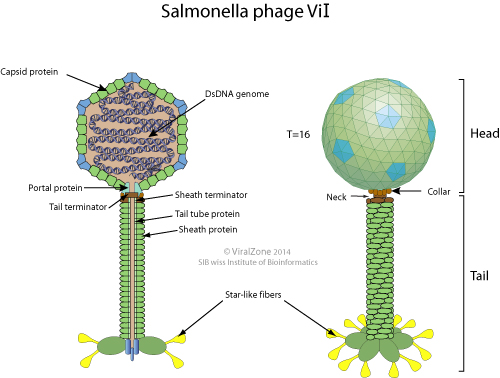
Summary[
edit] Description: English: Structure of a typical bacteriophage belonging to the Myoviridae family. The expanded inset shows a model of the gp37 tail fiber protein of phage T4 (PDB ID code 2XGF), visualized in Chimera (Pettersen et al., 2004), and VMD (Humphrey et al., 1996). Seven iron ions (red spheres) are coordinated octahedrally by histidine residues, forming a trimer as shown in the top image, while the bottom image shows a gp37 monomer. Pettersen, E. F., Goddard, T. D., Huang, C. C., Couch, G. S., Greenblatt, D. M., Meng, E. C., et al. (2004) "UCSF chimera–a visualization system for exploratory research and analysis". J. Comput. Chem., 25: 1605–1612.
doi:
10.1002/jcc.20084. Humphrey, W., Dalke, A., and Schulten, K. (1996) "VMD: visual molecular dynamics". J. Mol. Graphics, 14: 33–38.
doi:
10.1016/0263-7855(96)00018-5. Date: 25 May 2016, 19:42:01. Source:
[1]. Author: Chelsea Bonnain, Mya Breitbart and Kristen N. Buck.


.jpg)
.jpg)
.jpg)